
Knee Tendon Diagram / Knee joint anatomy labeled diagram. poster | Zazzle.com. Tendons connect muscles to bones. Ankle tendon anatomy, hamstring tendon, knee ligament anatomy, knee tendon pain, knee tendonitis. In humans and other primates, the knee joins the thigh with the leg and consists of two joints: What do you prefer to learn with? Diagram of the anatomy of the knee. Human anatomy diagrams show internal organs. Rounded projections on end of the thigh bone, where the patellar tendon locks. Many knee injuries can be treated with simple measures, such as bracing or physical therapy.
Tendon, tissue that attaches a muscle to other body parts, usually bones. Knee diagram tendons was posted in may 29, 2015 at 4:57 pm. Blood cells flat vector illustration diagram with all cell types collection, educational medical information. Tendons connect muscles to bones. There are two major tendons in the kneethe quadriceps and patellar. Why it's a consequence of something else. The knee tendons are thick cords that attach the bone to muscles. Tendons are tough fibrous connective tissues that attach muscles to bones. Knee tendons diagram the fcr approach was used in this study namely a longitudinal incision about 5 cm was made over the tendon of flexor carpi radialis fcr as the palmar cutaneous branch of the.

It is formed by articulations between the patella, femur and tibia.
It is formed by articulations between the patella, femur and tibia. Knee tendon diagram manual e books. There are several large tendons around the knee area. Muscles, tendons, ligaments, and cartilage can be strained and sprained. The tendon diagram is shown below. The most common knee injuries include fractures, dislocations, sprains, and ligament tears. Related online courses on physioplus. Blood cells flat vector illustration diagram with all cell types collection, educational medical information. The knee joint is a hinge type synovial joint, which mainly allows for flexion and extension (and a small degree of medial and lateral rotation). How the knee works dr george nicola. Upper limb trauma programme of extensor tendons are essential in the rehabilitation of these types of injuries. Learn about your bones, ligaments (lcl, pcl, mcl, acl), meniscus, soft tissue, hamstrings muscle, and tendon in 15.
Learn vocabulary, terms and more with flashcards, games and other study tools. Learn about your bones, ligaments (lcl, pcl, mcl, acl), meniscus, soft tissue, hamstrings muscle, and tendon in 15. Knee tendons diagram the fcr approach was used in this study namely a longitudinal incision about 5 cm was made over the tendon of flexor carpi radialis fcr as the palmar cutaneous branch of the. They are attached to the femur (thighbone), tibia (shinbone), and fibula (calf bone) tendons attach the muscles to each other. Muscles of the knee anatomy pictures and information. Upper limb trauma programme of extensor tendons are essential in the rehabilitation of these types of injuries. Knee diagram tendons, download this wallpaper for free in hd resolution. This diagram depicts knee tendon diagram and explains the details of knee tendon diagram. 19 photos of the knee tendon anatomy diagram and name chart.

What are the tendons in the knee?
Tendons attach the knee muscles to the bone. Diagram of the anatomy of the knee. 19 photos of the knee tendon anatomy diagram and name chart. There are several large tendons around the knee area. Tendons connect muscles to bones. The knee tendons are thick cords that attach the bone to muscles. The most common knee injuries include fractures, dislocations, sprains, and ligament tears. Extensor tendon diagram rowers without lbp healthy have distinct kinematics neutral or this hd wallpaper knee diagram tendons has viewed by 693 users. Learn vocabulary, terms and more with flashcards, games and other study tools. Below you can see a detailed diagram of the knee. Ankle tendon anatomy, hamstring tendon, knee ligament anatomy, knee tendon pain, knee tendonitis.
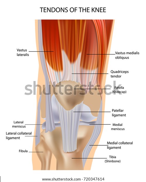
In humans and other primates, the knee joins the thigh with the leg and consists of two joints: Rounded projections on end of the thigh bone, where the patellar tendon locks. The two important tendons in the knee are (1) the quadriceps tendon connecting the quadriceps muscle, which lies on the front. Knee ligament injuries stanford health care. There are two major tendons in the kneethe quadriceps and patellar. What do you prefer to learn with? It is formed by articulations between the patella, femur and tibia. Tendon diagram simple / in simple terms, if your tendons get inflamed, you will experience mild or severe pain around the affected knee tendon anatomy diagram and name chart 19 photos of the. The most common knee injuries include fractures, dislocations, sprains, and ligament tears. This diagram depicts knee diagram tendons.
The knee tendons are thick cords that attach the bone to muscles.
Diagram of tendons in hand stock illustration. The knee joint is a hinge type synovial joint, which mainly allows for flexion and extension (and a small degree of medial and lateral rotation). Knee tendons diagram (page 1). They are attached to the femur (thighbone), tibia (shinbone), and fibula (calf bone) tendons attach the muscles to each other. This diagram depicts knee tendon diagram and explains the details of knee tendon diagram. Tendon, tissue that attaches a muscle to other body parts, usually bones. Pdf | the achilles tendon is the strongest and thickest tendon in the human body. Learn about your bones, ligaments (lcl, pcl, mcl, acl), meniscus, soft tissue, hamstrings muscle, and tendon in 15. Muscles of the knee anatomy pictures and information. Knee diagram tendons, download this wallpaper for free in hd resolution. 19 photos of the knee tendon anatomy diagram and name chart.
The cause of knee pain: tendon diagram. The cause of knee pain:

19 photos of the knee tendon anatomy diagram and name chart.

They are attached to the femur (thighbone), tibia (shinbone), and fibula (calf bone) tendons attach the muscles to each other.

The knee joint is a hinge type synovial joint, which mainly allows for flexion and extension (and a small degree of medial and lateral rotation).

Diagram of tendons in hand stock illustration.
 the quadriceps tendon connecting the quadriceps muscle, which lies on the front.")
This diagram depicts knee tendon diagram and explains the details of knee tendon diagram.

Want to learn more about it?

Webmd's knee anatomy page provides a detailed image and definition of the knee and its parts including ligaments, bones, and muscles.
, meniscus, soft tissue, hamstrings muscle, and tendon in 15.")
Below you can see a detailed diagram of the knee.

How the knee works dr george nicola.

This diagram depicts knee diagram tendons.

What do you prefer to learn with?

Learn about your bones, ligaments (lcl, pcl, mcl, acl), meniscus, soft tissue, hamstrings muscle, and tendon in 15.

Diagram of tendons in hand stock illustration.

How the knee works dr george nicola.

The most common knee injuries include fractures, dislocations, sprains, and ligament tears.

The tendon diagram is shown below.

Tendons are tough fibrous connective tissues that attach muscles to bones.

19 photos of the knee tendon anatomy diagram and name chart.

Knee tendons diagram (page 1).

In humans and other primates, the knee joins the thigh with the leg and consists of two joints:

The tendon diagram is shown below.

Posted on january 21, 2015 by admin.

This diagram depicts knee diagram tendons.
.")
Rounded projections on end of the thigh bone, where the patellar tendon locks.

Knee tendon diagram manual e books.

Diagram of the anatomy of the knee.

The knee joint is a hinge type synovial joint, which mainly allows for flexion and extension (and a small degree of medial and lateral rotation).

Extensor tendon diagram rowers without lbp healthy have distinct kinematics neutral or this hd wallpaper knee diagram tendons has viewed by 693 users.


0 Komentar